Items count : 18

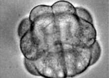
Embryonic development of Phallusia mammillata from 2 cell to gastrulation

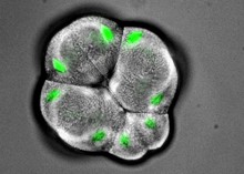
Interkinetic nuclear migration in early ascidian embryos

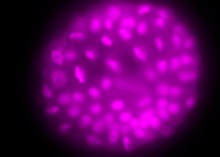
Waves of mitosis in the sea urchin embryo

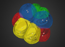
16 cell Vegetal 3D

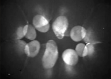
Development of the ctenophore embryo

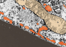
Section of the cortical vegetal region of the unfertilized egg of the ascidian Phallusia mammillata.

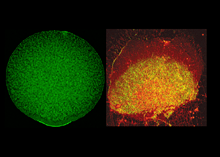
Just fertilized egg of the ascidian Phallusia mammillata (left) and corresponding vegetal pole cortex (right).


Gastrulation seen from the vegetal pole


110 cell stage seen from the vegetal pole


32 cell stage


Fertilized egg 5 minutes after sperm entry

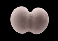
First cleavage 45 minutes after fertilization.

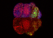
ERK1/2 activation in the developing marginal zone


Fertilized egg 5 minutes after sperm entry

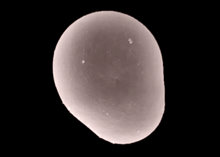
Fertilization triggers a microfilament-driven contraction.


The vegetal /contraction pole 10 minutes after fertilization


Phallusia embryo (16-cell stage during mitosis, vegetal view, posterior at bottom)


Phallusia embryo (64-cell stage during interphase, vegetal view, posterior on top)

